What Dental X-Rays Show Your Dentist
Bitewings, periapicals and 3D scans each show something different. What your dentist looks for, how often you need x-rays, and how low the dose is.
Dr. Edward J. Filangeri, DDS. Published
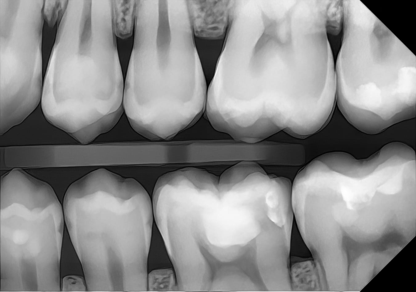
A dental exam shows the surfaces of your teeth and gums. X-rays show everything else: the spaces between teeth, the inside of old fillings, the roots, and the bone that holds it all in place. Here is what each type of x-ray is for.
Bitewings: between the teeth
Bitewings show the upper and lower back teeth together, from the crowns down to the top of the bone. They are the best way to find cavities that start between teeth, where neither you nor your dentist can see them. They also show decay under old fillings and early bone loss from gum disease.
Periapicals: the whole tooth
A periapical x-ray shows one or two teeth from tip to root, plus the surrounding bone. It is used to check a painful tooth, look for infection at the end of a root, follow up a root canal, or check an implant.
3D cone beam scans
For implant planning and some complex cases, a 3D cone beam scan shows the jaw in three dimensions, including the position of nerves and sinuses. It is not part of routine checkups.
How often you need them
There is no single schedule. The American Dental Association and the FDA recommend basing it on each patient’s risk. Someone who rarely gets cavities may need bitewings every year or two, while someone with active decay or gum disease may need them more often. Your dentist should be able to tell you why an x-ray is being taken.
How much radiation
Dental x-rays use a very low dose, and digital sensors need less than film did. Our office uses a wireless, direct-conversion digital sensor, which produces a sharp image at a low dose. If you are pregnant or might be, tell us before your visit.
See for yourself
You can zoom in on real digital x-rays, adjust the brightness and contrast, and see what Dr. Ed sees on our digital x-rays page.